
A diagnosis is built from the history and examination, not an image alone.
Knee condition guide
Knee Osteoarthritis
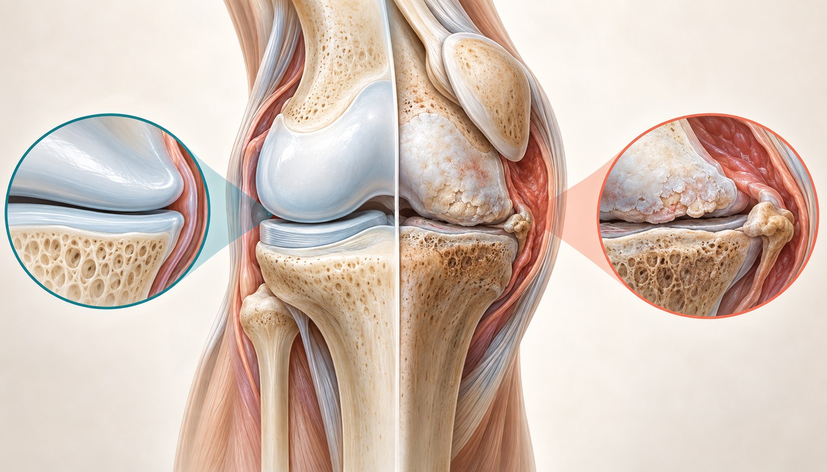
Knee osteoarthritis is a whole-joint condition. A useful evaluation connects your symptoms, function, examination, and imaging when needed, then matches care to the problem that is limiting you.
Quick orientation
What to know first
Symptoms and function guide care more than a single severity label.
Treatment choices can change as goals, health, and symptoms change.
What we may consider next
How we may treat this
These options depend on the diagnosis, exam findings, prior care, goals, and individual risk review. A listed pathway does not establish candidacy or guarantee a later procedure.
Establish first
Diagnostic or prognostic steps help clarify the pain generator or whether a later option fits.
Diagnostic / prognostic step
Genicular Nerve Block
May contribute to target assessment before a later nerve procedure; it does not establish RFA eligibility.
Options that may be considered
These are options, not required steps. Selection depends on the diagnosis and individual review.
Treatment option
Knee Joint Injection
May be considered for selected symptomatic knee osteoarthritis after diagnosis and option-specific review.
Treatment option
Platelet-Rich Plasma (PRP)
PRP may be discussed only after diagnosis-specific evidence, alternatives, and individual patient factors are reviewed.
May come later
A later procedure is considered only when evaluation and the response to earlier steps support it.
Later treatment option
Genicular Radiofrequency Ablation
Later therapeutic step only after diagnosis-specific evaluation and an appropriate target-assessment pathway.
Symptoms are clues, not a diagnosis
Common symptom patterns
People often describe activity-related aching, stiffness after rest, reduced motion, swelling, grinding, or difficulty with stairs, standing, walking, and getting up from a chair. Symptoms may vary from day to day. None of these features proves osteoarthritis by itself. Locking after an injury, marked instability, prolonged morning stiffness, or pain referred from the hip or spine can point toward a different or additional problem.
Activity-related ache or a deep joint pain
Brief stiffness after sitting or on first movement
Swelling, reduced bend or straightening, and a sense of fullness
Difficulty with stairs, walking distance, standing, or rising
Flares that temporarily change pain, sleep, and function
Inside the joint
A whole-joint process, not a simple wear meter
Age, prior injury, joint shape, alignment, muscle capacity, genetics, repetitive load, and metabolic health can influence risk. Osteoarthritis can involve the inner, outer, or kneecap-facing compartments in different combinations. Cartilage has limited repair capacity, but pain can also arise from bone, synovium, tendons, muscles, and sensitized nerves. That is why structural findings and day-to-day symptoms may not match neatly.
- Cartilage surface and joint-space change
- Bone remodeling and osteophytes
- Synovial irritation and intermittent fluid
- Meniscal degeneration and altered load distribution
- Strength, balance, sleep, and pain-processing influences
History first, imaging when it helps
How the diagnosis is evaluated
The visit starts with symptom timing, location, flares, injuries, prior care, walking tolerance, and the activities that matter to you. Examination may assess gait, alignment, swelling, motion, tenderness, strength, stability, hip and spine contribution, and neurovascular findings. Weight-bearing X-rays can clarify structure when they would change the decision. MRI, laboratory testing, ultrasound, or joint-fluid analysis is reserved for specific questions rather than used routinely.

History: pattern, duration, flares, function, injuries, and treatment response
Examination: motion, swelling, tenderness, strength, stability, gait, and nearby regions
Imaging: selected to answer a clinical question, not to replace the examination
Reassessment: response and function can refine the working diagnosis
Keep the differential open
Other causes a clinician may consider
Knee osteoarthritis can coexist with other problems. The differential changes with age, injury, swelling, locking, instability, fever, morning stiffness, nerve symptoms, and the location of pain. The goal is not to order every test. It is to identify findings that do not fit a routine osteoarthritis pattern and investigate those selectively.
- Meniscal or ligament injury
- Patellofemoral or tendon-related pain
- Bursitis or referred pain from the hip or spine
- Inflammatory or crystal arthritis
- Infection, fracture, tumor, or vascular disease when warning features are present
Core care
Build the non-procedural foundation
Education and individualized exercise are core care, not a test that must fail before something useful happens. Strengthening, aerobic activity, balance work, pacing, and supervised therapy can improve function. When relevant, weight-management support can reduce symptoms. Bracing, a cane, topical medication, or oral medication may help selected patients, but medication choice depends on kidney, stomach, heart, bleeding, and other health risks.
Progressive strengthening and aerobic activity
Physical therapy or supervised exercise when useful
Weight-management support when relevant
Brace, cane, pacing, sleep, and activity adjustments
Medication review with the appropriate clinician
A related option, not a requirement
Can weight management be part of knee osteoarthritis care?
For people with knee osteoarthritis who also have overweight or obesity, supported weight loss, particularly alongside exercise, has been shown in clinical trials to modestly improve pain and physical function. Weight management is not required to receive knee care and does not replace evaluation of the knee or other appropriate treatments.
Evidence at a glance
What the evidence can and cannot tell us
Exercise and weight management when relevant have broad guideline support. Joint corticosteroid injections have a short-term symptom role. Evidence for genicular radiofrequency treatment is technique dependent and includes both favorable and null sham-controlled trials. PRP guidance conflicts across professional groups, and preparation differences make studies difficult to compare. None of these options can promise a specific result for one patient.
Strongest agreement: education, exercise, and function-focused care
Short-term option: selected knee-joint injection
Conditional or limited option: genicular radiofrequency treatment
Conflicting option: PRP for selected patients
Unresolved: the best sequence and who will respond
Decision quality matters
Evidence, limits, and who it may fit
Guidelines summarize groups, while a treatment decision concerns one person. Selection should account for the diagnosis, compartment and pain pattern, symptom duration, function, prior response, medical risks, goals, cost, and willingness to continue active care. A procedure may reduce symptoms without changing joint structure. A reasonable plan defines what success would mean, how long to assess it, and what comes next if the response is incomplete.
- Confirm that osteoarthritis is the main pain generator
- Define a specific function or symptom goal
- Review contraindications and competing diagnoses
- Discuss evidence quality, alternatives, cost, and uncertainty
- Use a reassessment point rather than promise a duration
Referral and urgent signs
When another specialist may be the better next step
An orthopedic opinion can help when pain and loss of function remain substantial despite appropriate non-surgical care, or when deformity, instability, or advanced structural change affects the decision. Rheumatology may be appropriate for inflammatory features. Urgent or emergency assessment is more appropriate for a hot, rapidly swollen knee with fever, major trauma or deformity, inability to bear weight after injury, a newly locked knee, or symptoms suggesting a blood clot.
- Orthopedics for surgical decision-making
- Rheumatology for suspected inflammatory disease
- Urgent evaluation for infection, major trauma, or neurovascular concern
- Emergency care for chest pain, shortness of breath, or signs of a possible clot
The visit
What to expect from an evaluation
Bring a medication list, prior images or reports, and a short account of what the knee prevents you from doing. The first decision may be to refine the diagnosis, adjust exercise or medication strategy with the appropriate clinician, obtain targeted imaging, or discuss a procedure. You should leave knowing the working diagnosis, the purpose of the next step, realistic limits, alternatives, and how response will be measured.
A function-first history and focused examination
Review of prior care, medical risks, and imaging when relevant
A shared plan with a defined goal and reassessment point
Referral when another specialist is better suited to the problem
Common questions
Knee osteoarthritis FAQ
These answers are educational and cannot determine an individual diagnosis or treatment plan.
Does knee pain always mean osteoarthritis?
No. Meniscal or ligament injury, tendon or bursa irritation, inflammatory arthritis, crystal disease, infection, bone injury, nerve pain, and symptoms referred from the hip or spine can overlap. The history and examination help decide whether osteoarthritis is the main problem, one contributor, or not the likely explanation.
Do I need an MRI to diagnose knee osteoarthritis?
Usually not. Many cases can be assessed from the history and examination, with weight-bearing X-rays when structural information would change care. MRI may help when symptoms are atypical, an additional injury is suspected, or a specific treatment or surgical question cannot be answered another way.
Can an X-ray look severe when pain is mild?
Yes. Structural change and symptoms can be discordant in either direction. Images help describe the joint, but they do not measure your pain, walking tolerance, sleep, strength, or goals. Treatment decisions should combine the image with the clinical picture rather than follow the X-ray grade alone.
Will an injection rebuild the knee joint?
No injection should be described as proven to reverse osteoarthritis or rebuild cartilage. A corticosteroid injection may offer short-term symptom relief for some patients. PRP evidence and guidelines are mixed, products and protocols vary, and a large placebo-controlled trial did not show cartilage-volume preservation at 12 months.
Is a genicular nerve block the same as radiofrequency ablation?
No. A block places a short-acting medication near selected sensory targets and may provide temporary information about the pain pathway. Radiofrequency treatment uses thermal energy to alter signaling from selected nerves. A block response does not guarantee a particular radiofrequency outcome, and selection practices vary.
When should I discuss knee replacement with an orthopedic surgeon?
A surgical opinion may be appropriate when pain and functional loss remain substantial despite appropriate non-surgical care, or when deformity, instability, or structural disease changes the decision. Referral is an evaluation, not a commitment to surgery, and the timing should reflect health, goals, and expected benefit.
Diagnosis before procedure
Start with a clearer diagnosis
An evaluation can clarify whether osteoarthritis explains the pain and which conservative, interventional, or surgical pathway fits your goals.