
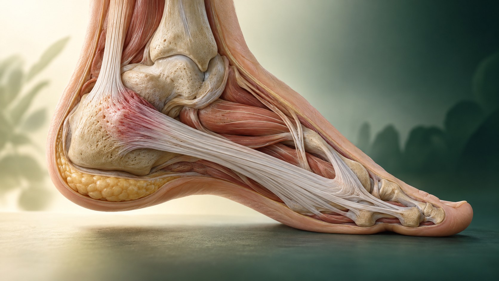
The plantar fascia supports the arch and is loaded with every step.
Leg, Ankle & Foot
Plantar Fasciitis
Plantar fasciitis commonly causes pain at the plantar-medial heel, especially with the first steps after rest. That pattern is useful, but heel pain can also come from bone, nerve, fat pad, tendon, or referred sources.
Quick orientation
What to know first
First-step pain is common, but it does not rule out other causes of heel pain.
Stretching, taping, footwear changes, and progressive loading usually come before injection.
What we may consider next
How we may treat this
These options depend on the diagnosis, exam findings, prior care, goals, and individual risk review. A listed pathway does not establish candidacy or guarantee a later procedure.
Options that may be considered
These are options, not required steps. Selection depends on the diagnosis and individual review.
Treatment option
Platelet-Rich Plasma (PRP)
PRP may be discussed only after diagnosis-specific evidence, alternatives, and individual patient factors are reviewed.
Symptoms are clues, not a diagnosis
What the pattern can feel like
Symptoms help define the pattern and show which daily, work, or athletic demands are difficult. They also guide the physical examination. No single symptom, tender spot, or self-test confirms the diagnosis, so a clinician still needs to consider nearby joints, nerves, referred pain, and other tissue problems.
Plantar-medial heel pain with the first steps after rest
Pain after prolonged standing or a rise in walking or running load
Tenderness near the medial calcaneal origin of the plantar fascia
Relevant anatomy
Anatomy and pain source
The plantar fascia is a strong band connecting the heel to the forefoot and helping support the arch. Plantar fasciitis usually reflects load-related fascial change near the heel origin rather than a dangerous inflammatory process.
History and examination first
How the diagnosis is evaluated
Evaluation uses the location and timing of pain, palpation, ankle and great-toe motion, walking and load history, and a neurologic screen. Imaging is not always needed, but ultrasound, radiographs, or MRI can help when the pattern is atypical or another diagnosis is suspected.

Keep the differential open
What else can look similar
A diagnosis is safer when the common alternatives are considered instead of treating pain location as proof.
- Calcaneal stress fracture
- Heel fat-pad pain
- Tarsal tunnel or another nerve problem
- Achilles or other posterior heel pathology
- Inflammatory or referred pain
Core care
Conservative care remains the foundation
Care should build capacity and reduce unnecessary protection while staying responsive to the exact diagnosis and symptom behavior.
Use plantar fascia-specific and calf stretching
Progress foot and calf loading
Consider taping, footwear changes, or orthoses for selected short-term needs
Use night splints when persistent first-step pain fits the pattern
Reassess atypical or non-improving pain
Evidence at a glance
Evidence at a glance
The evidence is not a scoreboard. Comparator choice, follow-up, patient selection, and the exact tissue problem can change the result.
The 2023 clinical guideline supports stretching and other targeted conservative strategies, with choices matched to the examination.
PRP-versus-corticosteroid reviews often favor PRP at three to six months, but trial methods and preparations vary.
That comparator evidence does not make PRP first-line and does not prove superiority to a complete rehabilitation pathway or placebo.
Decision quality matters
Evidence, limits, and who it may fit
Procedure consideration requires a confirmed diagnosis, correlation between symptoms and anatomy, review of prior care, contraindications, alternatives, goals, and the limits of the diagnosis-specific evidence. Relationship membership does not establish candidacy.
- The 2023 clinical guideline supports stretching and other targeted conservative strategies, with choices matched to the examination.
- PRP-versus-corticosteroid reviews often favor PRP at three to six months, but trial methods and preparations vary.
- That comparator evidence does not make PRP first-line and does not prove superiority to a complete rehabilitation pathway or placebo.
Referral and urgent signs
When another or urgent evaluation matters
Some findings should redirect the plan toward urgent care, another specialist, or a different diagnosis.
- Inability to bear weight after injury
- Marked swelling, bruising, fever, or an open wound
- Progressive numbness or weakness
- Night pain or systemic symptoms that do not fit a mechanical pattern
The visit
What to expect at an evaluation
The visit should narrow the diagnosis, clarify what has already been tried, and identify the next useful decision rather than defaulting to an injection. That may mean refining a rehabilitation plan, obtaining imaging only when it would change care, discussing a procedure with its evidence limits, or arranging another specialist evaluation.
Confirm the exact heel location and first-step pattern
Check ankle motion, arch and foot load, nerve findings, and stress-injury clues
Build conservative care first and reserve imaging or procedures for specific unanswered questions
Common questions
Plantar Fasciitis FAQ
These answers are educational and cannot determine an individual diagnosis or treatment plan.
Is a heel spur the cause?
A heel spur can appear on imaging in people with and without pain. The diagnosis depends more on the clinical pattern and examination than on a spur alone.
Do I need imaging?
Not always. Imaging becomes more useful when the pattern is atypical, symptoms persist, or a stress injury, nerve problem, tear, or another diagnosis is suspected.
Is PRP better than a steroid injection?
Some pooled comparisons favor PRP at later follow-up, but studies vary and often compare only those two injections. The result does not prove that PRP is best for every patient or that injection should come before rehabilitation.
Diagnosis before procedure
Start with a careful evaluation
The next step is to confirm the pain source, review prior care, and choose a plan that fits the diagnosis and your goals.