
Facet-related pain is a clinical hypothesis, not an imaging diagnosis.
Facet-mediated pain
Facet Joint Pain
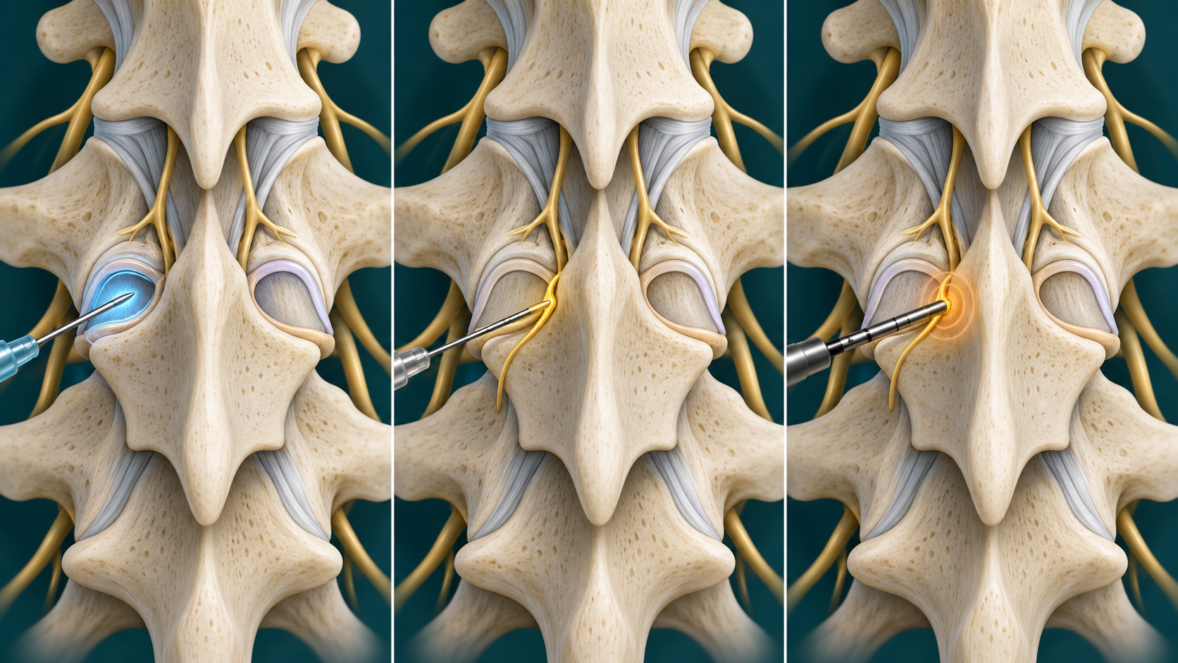
Facet joints guide motion at the back of the spine. They can contribute to axial neck or low-back pain, but common changes on a scan do not prove that a facet joint is the source.
Quick orientation
What to know first
History and examination help locate the problem but are not definitive.
A diagnostic block may inform a later decision; it does not obligate treatment.
What we may consider next
How we may treat this
These options depend on the diagnosis, exam findings, prior care, goals, and individual risk review. A listed pathway does not establish candidacy or guarantee a later procedure.
Establish first
Diagnostic or prognostic steps help clarify the pain generator or whether a later option fits.
Diagnostic / prognostic step
Medial Branch Blocks
This page explains the general diagnostic role. The cervical and lumbar guides explain region-specific anatomy and selection.
May come later
A later procedure is considered only when evaluation and the response to earlier steps support it.
Later treatment option
Radiofrequency Ablation
This page explains the general later-treatment role. The cervical and lumbar guides explain region-specific anatomy and selection.
Alternatives, not a sequence
These options may be considered separately; one does not automatically lead to another.
Selected alternative
Facet Joint Injection
Alternative facet intervention context without replacing regional diagnosis and treatment pages.
Choose the guide that matches your question
Explore the specific guides.
Use these links to learn about related symptoms, conditions, and treatments. An individual evaluation is still needed to determine a diagnosis or whether a treatment may be appropriate.
Clinical guide
Cervical Facet Joint Pain
Region-specific cervical facet-mediated pain, diagnostic medial branch block considerations, and possible RFA pathway.
Open this guideClinical guide
Lumbar Facet Joint Pain
Region-specific lumbar facet-mediated pain, diagnostic medial branch block considerations, and possible RFA pathway.
Open this guideSymptoms are clues, not a diagnosis
What the pattern can feel like
Symptoms can help define a pattern, but they do not confirm which structure is responsible. Distribution, timing, aggravating factors, neurologic findings, function, and important alternatives all shape the evaluation. A familiar pattern can still have more than one contributor, and a change in pattern may require a different assessment.
Pain is often axial rather than dominated by limb symptoms.
Movement, posture, or sustained loading may change symptoms.
The pattern overlaps with disc, muscle, sacroiliac, nerve-root, and other pain sources.
Relevant anatomy
Anatomy and pain mechanisms
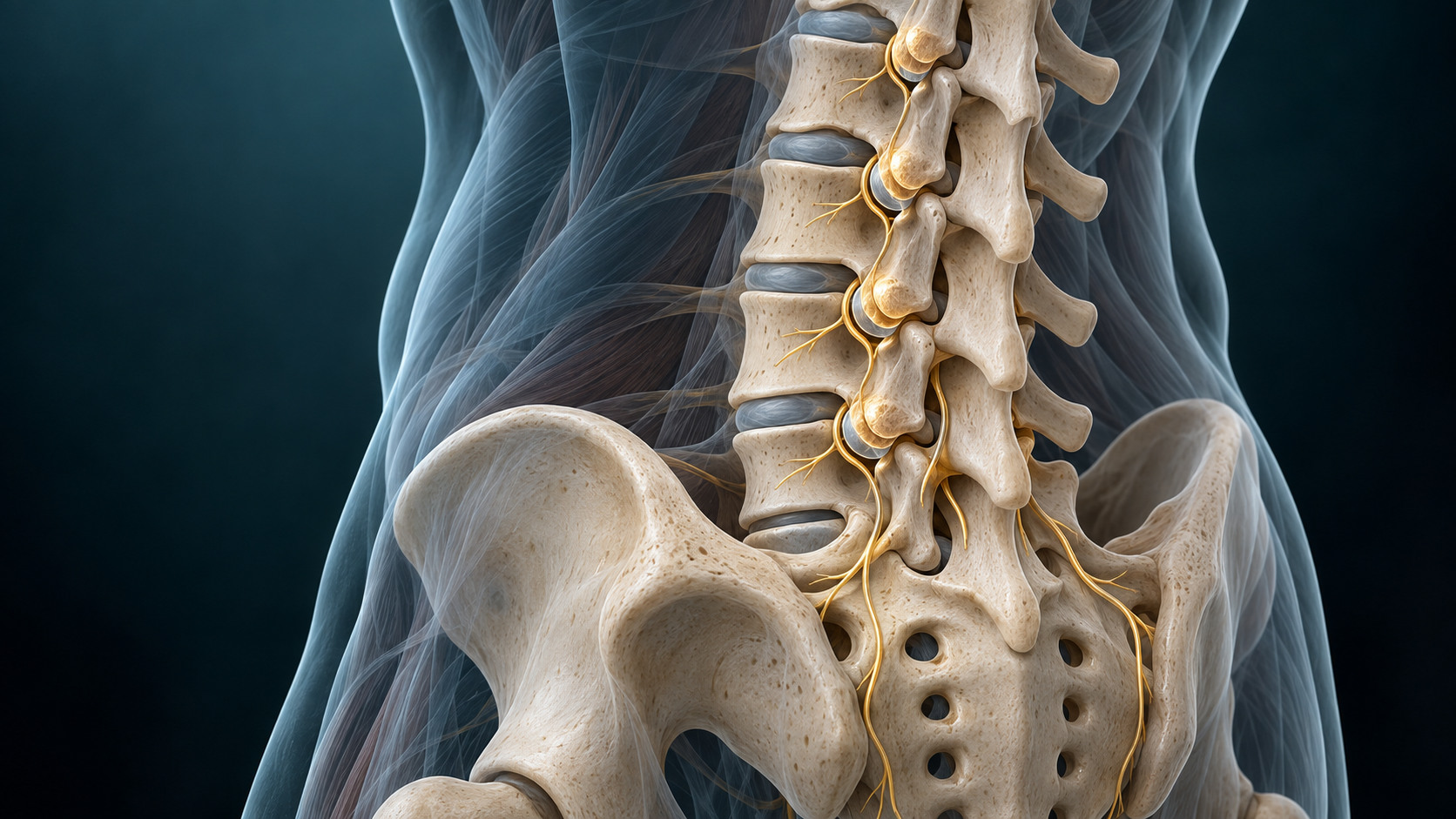
Each spinal level has paired facet joints supplied by small medial branch nerves. Cervical and lumbar anatomy, symptoms, risks, and procedure technique differ, so region-specific pages carry those details.
History and examination first
How the diagnosis is evaluated
Evaluation starts with history, examination, neurologic screening, and review of prior care. Imaging may identify degeneration or exclude another concern, but common facet changes can also appear in people without pain. A carefully performed diagnostic block may be considered when the result would change management.
Keep the differential open
What else can look similar
Keep common and serious alternatives open until the evidence supports a narrower conclusion.
Disc-related pain
Myofascial pain
Sacroiliac pain
Radiculopathy or spinal stenosis
Fracture, infection, inflammatory disease, or another urgent cause when clinical features suggest it
Core care
Conservative and coordinated care
Procedure discussions belong inside a broader plan that protects function.
Education and paced activity
Exercise or physical therapy matched to the diagnosis
Medication review and risk reduction
Reassessment when the pattern changes or does not respond as expected
Evidence at a glance
Evidence at a glance
Evidence strength, comparator, follow-up, and selection affect interpretation.
Imaging findings correlate poorly with a painful facet source.
Consensus recommendations differ on selection thresholds and block protocols.
Recent placebo-controlled syntheses report small or uncertain average benefits, while selected-patient reviews report more favorable RFA outcomes.
Decision quality matters
Evidence, limits, and who it may fit
A listed option does not establish candidacy. Diagnosis, anatomy, prior care, risk, alternatives, goals, and evidence must align.
Referral and urgent signs
When urgent or different evaluation matters
These findings can change the timing or destination of care.
New or progressive weakness
Bowel or bladder dysfunction or saddle numbness
Fever, unexplained weight loss, major trauma, cancer history, or rapidly worsening pain
The visit
What to expect at an evaluation
The visit should narrow the diagnosis, identify safety and referral needs, review imaging and prior care in context, and choose the next useful decision. The clinician should explain uncertainty, distinguish clinical evidence from coverage rules, and define what information a test or treatment is expected to add before it is performed.
Review the pain pattern, neurologic symptoms, imaging, and prior care
Set measurable pain and functional goals before any procedure
Choose the next step from the full clinical picture rather than one isolated test
Common questions
Facet Joint Pain FAQ
These answers are educational and cannot determine an individual diagnosis or treatment plan.
Does arthritis on a scan prove facet pain?
No. Facet changes are common and may not match symptoms.
Are cervical and lumbar facet pain the same?
They share a joint and nerve concept, but anatomy, differential diagnosis, risks, and technique differ.
Does a positive block mean RFA is required?
No. A block is one part of an individualized decision.
Diagnosis before procedure
Start with a careful evaluation
A focused evaluation can clarify the diagnosis, important alternatives, and the next step that fits the clinical picture.