
Symptoms, examination, and imaging must be interpreted together.
Spine and nerve conditions
Lumbar Spinal Stenosis
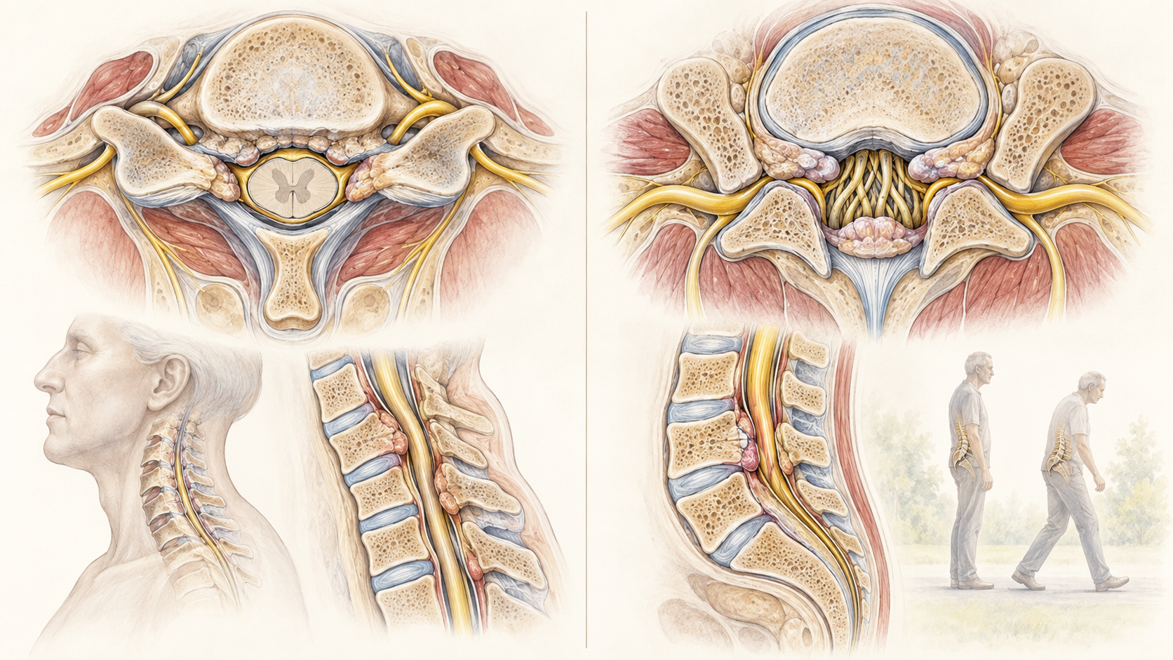
Lumbar spinal stenosis is narrowing that may crowd nerves in the lower spine. A characteristic pattern is leg discomfort or heaviness with standing or walking that improves with sitting or bending forward, but imaging alone does not establish the syndrome.
Quick orientation
What to know first
A scan finding can be relevant without being the entire diagnosis.
Progressive neurologic or cord-related findings can change the urgency and destination of care.
What we may consider next
How we may treat this
These options depend on the diagnosis, exam findings, prior care, goals, and individual risk review. A listed pathway does not establish candidacy or guarantee a later procedure.
Options that may be considered
These are options, not required steps. Selection depends on the diagnosis and individual review.
Treatment option
Lumbar Epidural Steroid Injection
May be considered for selected radicular symptoms; it does not treat every stenosis presentation.
Treatment option
Minuteman Interspinous-Interlaminar Fusion
May be considered only for the procedure's confirmed selection criteria after structural, neurologic, and surgical review.
Alternatives, not a sequence
These options may be considered separately; one does not automatically lead to another.
Selected alternative
Caudal Epidural Steroid Injection
Alternative access pathway when target, anatomy, prior surgery, and symptom pattern support it.
Symptoms are clues, not a diagnosis
What the pattern can feel like
Symptoms help define a syndrome, but they do not confirm which structure is responsible. Distribution, timing, aggravating factors, neurologic findings, function, and important alternatives all shape the evaluation.
Pain may be only one part of the pattern.
Numbness, tingling, weakness, balance, walking tolerance, and function help define the problem.
Symptoms can overlap with more than one spinal and non-spinal condition.
Relevant anatomy
Anatomy and pain mechanisms
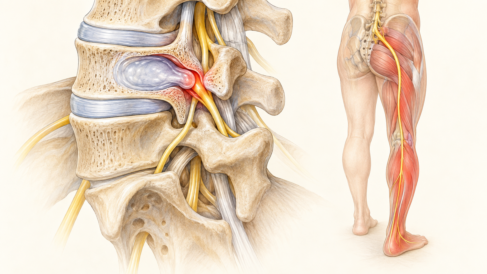
The lumbar canal and nerve-exit openings can narrow from age-related changes in discs, joints, and ligaments. Symptoms may affect both legs, walking tolerance, or a single nerve root, and the pattern determines which options are relevant.
History and examination first
How the diagnosis is evaluated
Evaluation begins with the time course, symptom distribution, neurologic examination, functional effects, and urgent features. Imaging can identify anatomy and important alternatives, but the result must correlate with the clinical pattern.
Keep the differential open
What else can look similar
Keep common and serious alternatives open until the evidence supports a narrower conclusion.
Axial neck or low-back pain without radiculopathy
Peripheral nerve, joint, vascular, or other non-spinal sources
Facet, disc, stenosis, or nerve-root disorders with overlapping features
Infection, fracture, tumor, inflammatory disease, or another urgent cause when the history suggests it
Core care
Conservative and coordinated care
Procedure discussions belong inside a broader plan that protects neurologic function and daily activity.
Education and paced movement
Exercise or physical therapy tailored to the diagnosis
Medication review and risk reduction
Reassessment for changing neurologic findings or function
Evidence at a glance
Evidence at a glance
Evidence strength, comparator, follow-up, and selection affect interpretation.
Imaging severity and symptom severity do not always match.
Exercise and multimodal rehabilitation can improve function for some people with neurogenic claudication.
Epidural steroid evidence for lumbar stenosis is limited and inconsistent, with no reliable short-term pain advantage established.
Decision quality matters
Evidence, limits, and who it may fit
A listed option does not establish candidacy. Diagnosis, anatomy, prior care, risk, alternatives, goals, and evidence must align.
Referral and urgent signs
When urgent or different evaluation matters
These findings can change the timing or destination of care.
New or progressive weakness
New bowel or bladder dysfunction or saddle numbness
Fever, major trauma, cancer history, or rapidly worsening pain
The visit
What to expect at an evaluation
The visit should narrow the diagnosis, identify safety and referral needs, review imaging and prior care in context, and choose the next useful decision. The clinician should explain uncertainty and what a treatment is expected to add before it is performed.
Confirm the symptom pattern and neurologic examination
Review imaging only in the context of symptoms and examination
Choose among rehabilitation, medication, watchful reassessment, procedure discussion, or referral based on the complete picture
Common questions
Lumbar Spinal Stenosis FAQ
These answers are educational and cannot determine an individual diagnosis or treatment plan.
Is this the same as sciatica?
No. Stenosis can cause radicular symptoms, but neurogenic claudication more often relates to standing and walking tolerance.
Can circulation problems mimic lumbar stenosis?
Yes. Vascular claudication and other non-spinal causes of walking-limited leg symptoms must remain in the differential.
Do injections reverse stenosis?
No. An injection does not widen the canal and evidence for stenosis-associated symptoms is limited.
Diagnosis before procedure
Start with a careful evaluation
A focused evaluation can clarify the diagnosis, important alternatives, and the next step that fits the clinical picture.