
Symptoms, examination, function, and imaging must be interpreted together.
Joint and musculoskeletal conditions
Sacroiliac Joint Pain
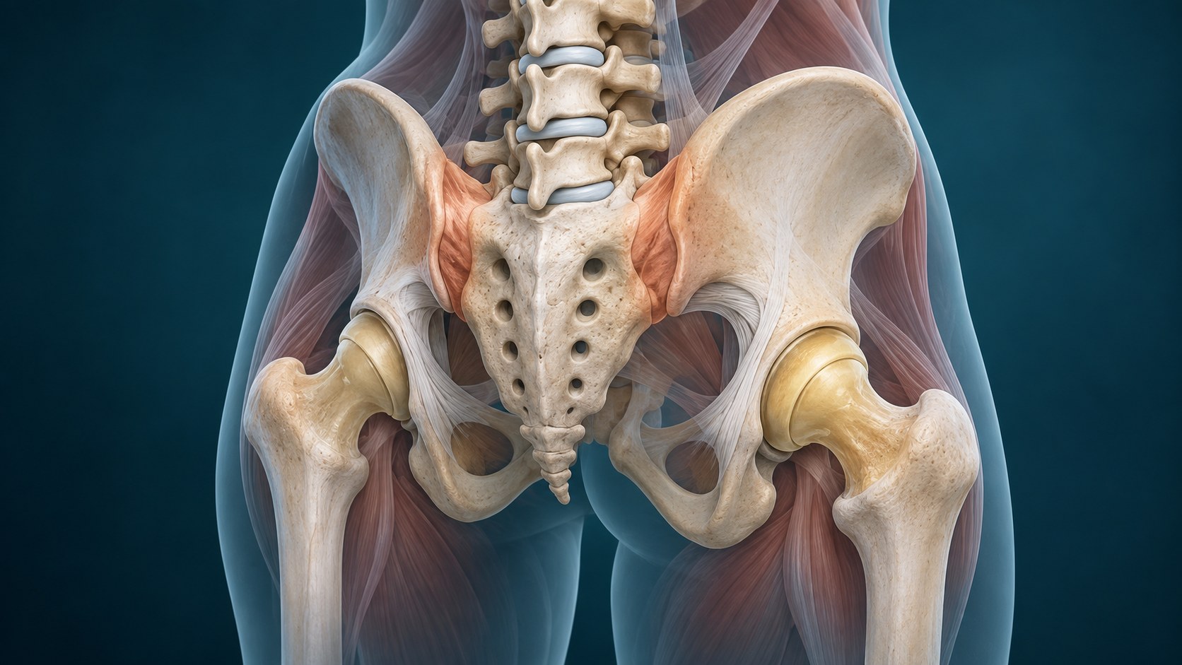
Pain near the back of the pelvis can come from the sacroiliac joint complex, but location or imaging alone cannot confirm the source. Diagnosis depends on the full pattern and how competing causes are addressed.
Quick orientation
What to know first
Neighboring joints, tendons, nerves, and referred pain can overlap.
A procedure is one possible tool, not proof of the diagnosis or an automatic next step.
What we may consider next
How we may treat this
These options depend on the diagnosis, exam findings, prior care, goals, and individual risk review. A listed pathway does not establish candidacy or guarantee a later procedure.
Establish first
Diagnostic or prognostic steps help clarify the pain generator or whether a later option fits.
Diagnostic / prognostic step
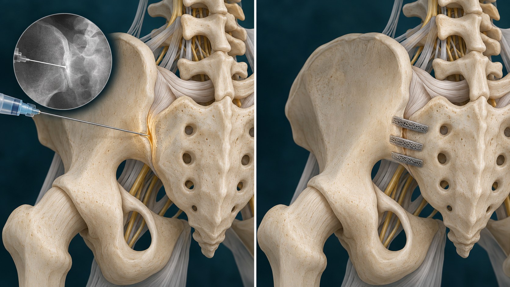
Sacroiliac Joint Injection
May contribute to a diagnosis-specific injection pathway and selected therapeutic assessment.
May come later
A later procedure is considered only when evaluation and the response to earlier steps support it.
Later treatment option
Posterior Sacroiliac Joint Fusion
Later treatment pathway only after diagnosis confirmation, conservative care, and procedure-specific selection.
Symptoms are clues, not a diagnosis
What the pattern can feel like
Symptoms help define a syndrome, but they do not confirm which structure is responsible. Distribution, timing, aggravating factors, neurologic findings, function, and important alternatives all shape the evaluation.
Pain location and movement sensitivity can narrow the differential but rarely identify one structure alone.
Stiffness, range of motion, loading tolerance, weakness, swelling, and daily function add diagnostic context.
The pattern should be checked for referred pain, neurologic features, inflammatory disease, injury, and other alternatives.
Relevant anatomy
Anatomy and pain mechanisms
The SI joint complex includes the joint surfaces, capsule, and strong surrounding ligaments between the sacrum and pelvis. Intra-articular and extra-articular structures may contribute, while lumbar and hip disorders can create a similar pattern.
History and examination first
How the diagnosis is evaluated
Evaluation combines the history, movement and loading pattern, focused examination, functional goals, and selective imaging. Structural change can support a diagnosis, but imaging severity and symptom severity may not match.
Keep the differential open
What else can look similar
Keep common and serious alternatives open until the evidence supports a narrower conclusion.
Referred spinal or nerve-related pain
Tendon, bursa, muscle, or neighboring joint disorders
Inflammatory arthritis, infection, fracture, or another less common cause when the history suggests it
Core care
Conservative and coordinated care
Procedure discussions belong inside a broader plan that protects neurologic function and daily activity.
Education, pacing, and movement matched to irritability
Progressive strength, mobility, and function when appropriate
Medication review that accounts for individual risks
Reassessment when the pattern changes or improvement stalls
Evidence at a glance
Evidence at a glance
Evidence strength, comparator, follow-up, and selection affect interpretation.
Provocation-test clusters may help rule the SI joint out more confidently than they can rule it in.
Image-guided diagnostic injections can add information but are imperfect and are interpreted with the complete clinical picture.
Evidence for treatment pathways varies, and fusion evidence remains limited and conflicting.
Decision quality matters
Evidence, limits, and who it may fit
A listed option does not establish candidacy. Diagnosis, anatomy, prior care, risk, alternatives, goals, and evidence must align.
Referral and urgent signs
When urgent or different evaluation matters
These findings can change the timing or destination of care.
Fever, a hot swollen joint, or systemic illness
Major trauma or inability to bear weight or use the limb
Rapidly progressive weakness, numbness, or a new emergency symptom pattern
The visit
What to expect at an evaluation
The visit should narrow the diagnosis, identify safety and referral needs, review imaging and prior care in context, and choose the next useful decision. The clinician should explain uncertainty and what a treatment is expected to add before it is performed.
Define the symptom pattern and meaningful functional limits
Examine the joint and neighboring regions before attributing symptoms to one structure
Review imaging only in clinical context
Choose among rehabilitation, medication review, a selected procedure discussion, or referral without assuming a fixed sequence
Common questions
Sacroiliac Joint Pain FAQ
These answers are educational and cannot determine an individual diagnosis or treatment plan.
Is SI joint pain the same as low-back pain?
No. It can overlap with low-back and hip symptoms, so the source must be evaluated rather than assumed from location.
Does an MRI prove the SI joint is painful?
No. Imaging can identify anatomy and alternatives but does not establish the symptomatic structure by itself.
Does a helpful injection mean fusion is next?
No. An injection result is one part of a broader assessment and does not create an automatic surgical pathway.
Diagnosis before procedure
Start with a careful evaluation
A focused evaluation can clarify the diagnosis, important alternatives, and the next step that fits the clinical picture.